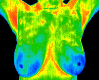
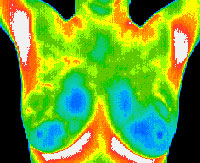
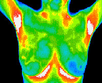
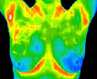
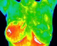
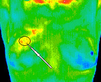
What is Digital Infrared Thermography Imaging, and how can it benefit me?
Simply put, Digital Infrared Thermography is a 15 minute painless and non invasive test of physiology. It is a valuable method available for visualizing pain and physiology. A special camera is used to provide instant thermal images or “thermograms” of the whole body or just areas being investigated. The digitized images are stored on a computer and sent electronically to a Medical Thermologist for interpretation and analysis.
This valuable procedure offers earlier detection of Breast Disease as an adjunct to self examination, doctor examination, and mammography alone. Digital Infrared Thermography detects the subtle physiologic changes that accompany breast pathology, whether it is cancer, fibrocystic disease, an infection or a vascular disease. With this, your doctor can plan accordingly and lay out a careful program to further diagnose or monitor you during and after treatment.
The Best Breast Test: The Promise of Thermography
Christine Northrup MD, Board Certified OB/GYN and author of acclaimed books on womens health, is a advocate of Medical Thermography: Huffington Post Healthy Living:
Read more on Christiane Northrup MD’s thoughts about the benefits of breast thermography and the importance of early detection!
As an example . . . If you had a penny inserted into your breast, your body would react by sending white blood cells and histamine, causing an inflammatory reaction. A mammogram would JUST just see the penny; a thermogram would see the temperature of the penny and the reaction to the penny…not the penny. When cancer cells start to grow, these cells recruit a blood supply to feed them. This is called angiogenesis. Angiogenesis in breast tissue is thought to be a sign that breast cancer may follow. Thermography picks up a thermal pattern from that blood supply, often before a mammogram can detect the cells.
In addition to its benefits in early detection of Breast Disease, this clinical imaging procedure detects and monitors a number of diseases and physical injuries, by showing the thermal abnormalities present in the body, including:
 Back injuries Digestive Disorders
Arthritis Carpal Tunnel Syndrome
Headache Disc Disease
Nerve damage Inflammatory Pain
Unexplained Pain Skin Cancer
Fibromyalgia Referred Pain Syndrome
RSD (CRPS) Sprain/Strain
Dental and TMJ Whiplash
Artery Inflammation Stroke Screening
Vascular Disease
Here are some frequently asked questions about Digital Infrared Thermography:
Q. How does Digital Infrared Thermography Work?
A. Medical Digital Infrared Imaging is a noninvasive diagnostic technique that allows the examiner to visualize and quantify changes in skin temperature. An infrared scanning device is used to convert infrared radiation emitted from the skin surface into electrical images that are converted to color on a monitor. This visual image graphically maps the body temperature and is referred to as a thermogram. The spectrum of colors indicate an increase or decrease in the amount of infrared radiation being emitted from the body surface. Since there is a high degree of thermal symmetry in the normal body, subtle abnormal temperature asymmetries can be easily identified.
Q. How does Digital Infrared Thermography compare to an X-ray, C.T., Ultrasound or M.R.I.?
A. X-ray, C.T., Ultrasound and M.R.I. are tests of anatomy and structure. Digital Infrared Thermography is unique in it’s capability to show physiological change and metabolic processes. It has also proven to be a very useful complementary procedure to other diagnostic methods.
Q. What is its clinical value?
A. Its value is to define the extent of a lesion of which a diagnosis has previously been made; localize an abnormal area not previously identified, so further diagnostic tests can be performed; detect early lesions before they are clinically evident; and monitor the healing process.
Q. What is the difference between a Thermogram and a Mammogram?
A. A Thermogram is a test of physiology, a Mammogram one of structure. In a thermogram you will see temperature and a thermal pattern. A mammogram indicates densities, sizes and shapes.
Q. What is the benefit of a test that shows physiology?
A. Cellular changes can create an abnormal thermal pattern and can often be viewed by a Thermogram before a lesion is dense enough for a Mammogram.
Q. How are you certified?
A. Thermographic technicians are trained and certified by the American College of Clinical Thermology (formerly hosted at Duke University).
Q. Who reads the images and reports?
A. Images are analyzed by medical doctors, who are all board certified Thermologists, certified by the American College of Clinical Thermology.
Q. How quickly will I get my report back?
A. Reports are normally ready within 48 hours.
Q. What is the difference between grayscale and color Thermograms?
A. There’s no difference in resolution between color and grayscale with modern digitized images.
Q. How important is “high definition” Thermography?
A. Describing a Thermogram as “high definition” may be confusing and misleading, because most high-definition images are produced by software manipulation of the data. The higher the definition, the better the picture will look, but this does not mean that the accuracy is any better. The actual definition is not as important as how accurate and sensitive those temperature measurements are.
Q. Why do I need to come back in three months for another breast scan?
A. Before any changes can be evaluated, an accurate and stable baseline must be established. A baseline cannot be determined with only one scan, since there would be no way of knowing if it is a normal pattern, or if it is actually changing at the time of the first exam.

 Back injuries Digestive Disorders